animal cell under microscope diagram
A typical animal cell is 1020 μm in diameter which is about one-fifth the size of the smallest particle visible to the naked eye. Students will observe onion cells under a microscope.
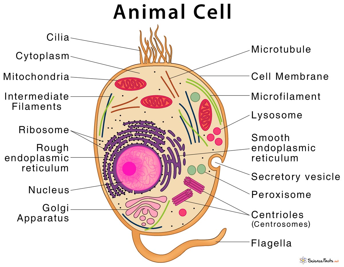
Animal Cell Structure Parts Functions Types With Diagram
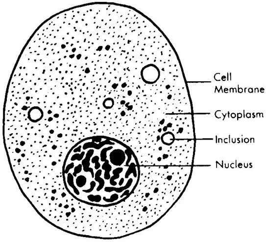
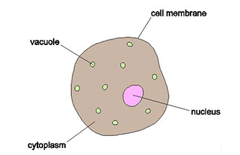
The living material between the nucleus and the cell surface.
. Ad Over 27000 video lessons and other resources youre guaranteed to find what you need. A sample picture of each a plant animal and. An average mammal cell has a diameter of 1020 m or around one-fifth the size of the smallest particle discernible with the unaided eye.
It is a rigid layer that is composed of cellulose glycoproteins lignin pectin and hemicellulose. Pin on R Cell Assignment. Simple squamous epithelium under a microscope consists of a single layer of thin flat and scale-like cells.
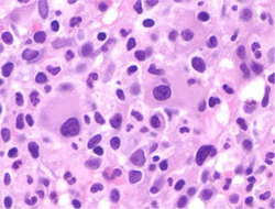
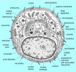
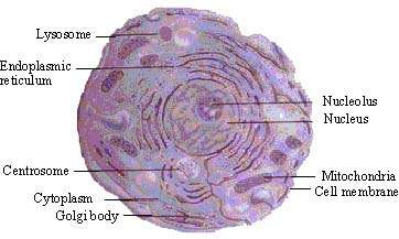
It was not until good light microscopes became available in. The first animal cell was observed under an optical microscope which clearly showed the nucleus and microfilament network in red and blue colors respectively. In the cell there are organelles which are suspended within an aqeuos medium and contained within plasmamembrane.
Diagram of parts of a microscope. Students will discover that their skin is made up of cells. Just under the rigid cell wall is the more fluid cell membrane.
It is located outside the cell membrane and is completely permeable. Print the scripts for animal and plant cells. Students will discover that onions are made up of cells.
Animal cell under the microscope. These cells are joined together by an intercellular junction and rest on the. General Microscope Handling Instructions Hold with one hand under the base and other hand on the C-shaped arm to bring the microscope.
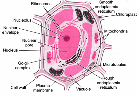
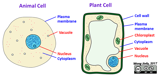
When looking under a microscope the cell wall is an easy feature to distinguish plant cells. Typical Animal Cell Pinocytotic vesicle Lysosome Golgi vesicles Golgi vesicles rough ER endoplasmic reticulum Smooth ER no ribosomes Cell plasma membrane. Diagram Of Animal Cell Under Microscope.
Animal Cell Under Light Microscope.
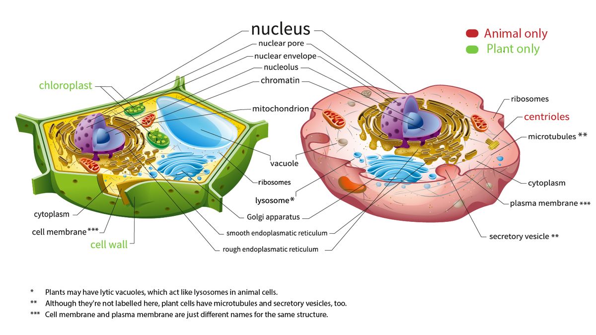
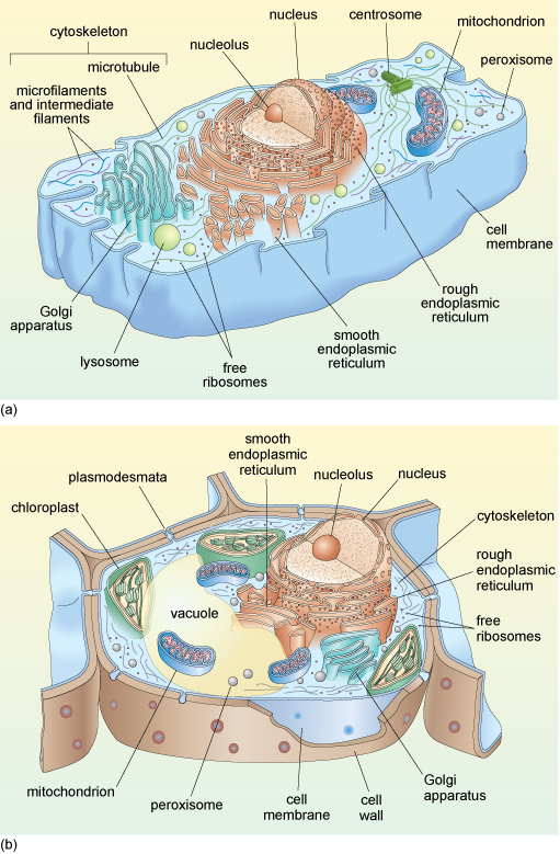
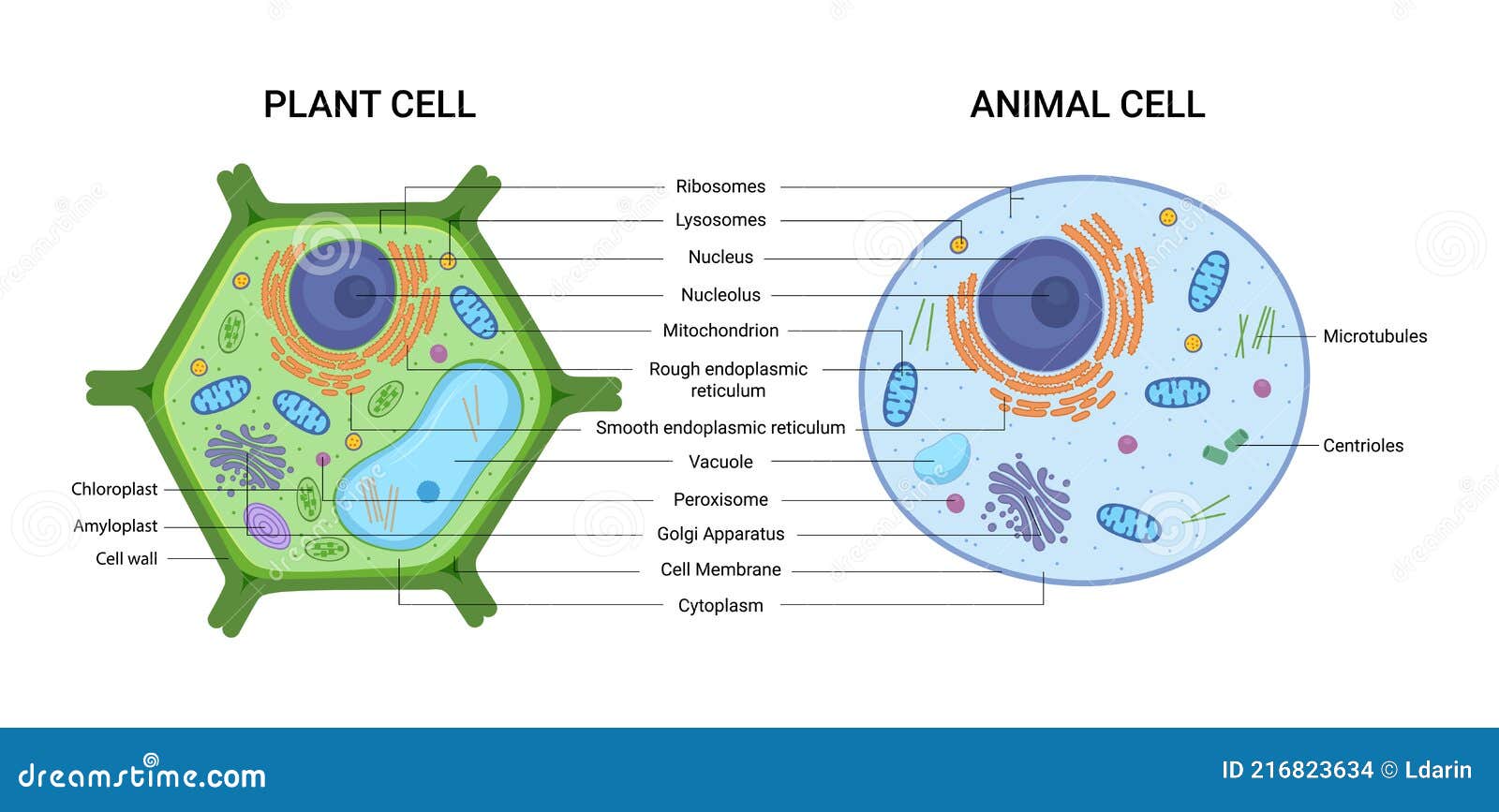
Here S How Plant And Animal Cells Are Different Howstuffworks
Animal Cells And Plant Cells Cell Processes

50 Microscope Slide High Scale Magnification Magnification Animal Cell Stock Photos Pictures Royalty Free Images Istock
Cell Structure Article About Cell Structure By The Free Dictionary
Cape Biology And Chemistry Syllabus Biology Module 1 2 Cell Structure 2 1 Make Drawings Of Typical Animal And Plant Cells As Seen Under The Light Microscope

1 2 Difference Between Plant And Animal Cells Cells As The Basic Units Of Life Siyavula
Pinkmonkey Com Biology Study Guide Chapter 3 Cell The Basic Unit Of Life
A Tour Of The Cell View As Single Page

Animal Cell Diagram By Russell Kightley
Animal Plant Cells Stock Illustrations 374 Animal Plant Cells Stock Illustrations Vectors Clipart Dreamstime

Illustrate Only A Plant Cell As Seen Under Electron Microscope How Is It Different From Animal Cell

Structure Of Animal Cell And Plant Cell Under Microscope Diagrams

Animal Cells Under Microscope Stock Photo Picture And Royalty Free Image Image 57823181
Illustrate Only A Plant Cell As Seen Under Electron Microscope How Is It Different From Animal Cell Cbse Class 9 Science Learn Cbse Forum
Olcreate Tessa Btw Module 1 Secondary Science Biology Resource 1 Background Information On Cells

Plant Cell Under The Microscope 2 Plant Cell Things Under A Microscope Plant Cell Picture
Cells Mr Wong S Class Website
What Are The Visible Plant Animal Cell Organs On Light Microscope Quora
What Is The Correct Diagram Of Plant And Animal Cell Quora